The Trabecular Bone Pattern as a window into Osteoporosis
This is the work from my master’s thesis. I never published it in a journal, so I’ve posted it here hoping it might be useful to others.
What is Trabecular Bone?
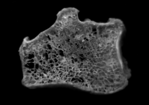
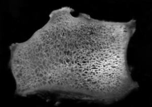
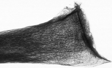
When you x-ray a bone, you often see a pattern (Figure 1) caused the trabecular network inside the core of the bone. You can see the trabecular network directly by looking at vertical slices through two bones; Figures 2 and 3 show photos of bone slices of someone with and someone without osteopenia (the thinning of bone that leads to osteoporosis). The trabeculae themselves are very different, much thicker and more numerous for the subject without osteopenia.
If the pattern on the x-ray changes with the progression of osteopenia, we could we could test for osteopenia using standard x-ray equipment, rather than needing specialized equipment like a DEXA scanner.

Figure 2: Section through the radius bone of a person with osteopenia, showing the thin trabecular network.

Figure 3: Section through the radius bone of a person without osteopenia, showing the dense trabecular network.
Analyzing the Bone pattern
Can osteopenia, the thinning of bone that leads to osteoporosis, be measured by changes in the trabecular pattern on an x-ray? This is the question I set out to investigate in my MSc thesis. Here’s the abstract:
This project is concerned with the extraction of information relating to the trabecular structure of bones from planar x-ray radiographs, with a view to gaining better information about osteopenia. Current clinical assessment of bone mineral density (BMD) ignores structural information.
High resolution planar x-ray radiographs were taken of five human radii excised from cadavers with a range of age, sex and build. BMD values for the radii were obtained using a DEXA scanner and the bones were sliced to obtain trabecular structure scores. Image processing techniques were applied to regions of the digitized x-ray images showing the radiographic trabecular pattern of the distal radius. Techniques similar to those used by Geraets et al. in previous studies were found to produce parameters correlating highly with BMD and less highly with trabecular structure scores. Image signal to noise ratio (SNR) was found to be closely related to BMD. High dependence of the parameters on image SNR was thought to explain the correlations previously found with BMD. The parameters are thought to be dependent on trabecular structure only via the BMD.
New measures were developed which appear to be less dependent on image SNR. Spectral analysis of the images yeilded information which correlated well with both BMD and structural scores. A new measure analyzing the skeletonized image was found to correlate more highly with trabecular structure scores than with BMD. This may potentially provide a way of analyzing the progression of diseases such as osteopenia using ordinary planar x-ray radiographs and obviate the need for specialized scanners.
You can download the full thesis here: Analysis of the radiographic trabecular pattern of the ultradistal radius (4MB pdf).
Can the trabecular bone pattern reveal osteopenia?
So, can osteopenia be measured by changes in the trabecular pattern on an x-ray?
Yes, but we have to be careful.
Most of the bone density is contributed by the cortex—the thick bone at the outside. As this increases the trabecular bone pattern stands out less on the x-ray image because it’s signal to noise ratio (SNR) decreases. The SNR of the x-ray pattern therefore correlates well with the overall bone density, and pattern measures that are sensitive to noise therefore correlate well with bone density. This explained why some previously used measured correlated with bone density, but the bad news is that we can’t use them to measure osteopenia because when a greater thickness of flesh is x-rayed, this also drops the SNR of the pattern, confounding the result. These measures therefore can’t substitute for a DEXA scanner, which ratios absorption of different x-ray wavelengths to parse out the contribution of flesh and contribution of bone.
Filtering the images to remove noise can eliminate the SNR effect. Interestingly, different measures that look at the connectedness of the trabecular pattern on these filtered images correlate well with the bone density, suggesting that, in principle, this technique should work.
References
Analysis of the radiographic trabecular pattern of the ultradistal radius (1996) Dayel MJ. A thesis presented to the Centre for Biological and Medical Systems, Imperial College, London in partial fulfillment of the requirements for the degree Master of Science in Engineering and Physical Science in Medicine

I am beginning my doctoral work on bone density in bottlenose dolphins. One of the intial studies we are looking at conducting is to compare the density in different regions (~2 cm diameter) among the radius and ulna to define the range/gradient of densities observed throughout the areas of the bone. Was hoping you may be interested in providing any insight to this and other questions we are looking at pursuing.
Thank you for your time
-James